Biopsy and histopathology: A microscopic view to identifying disease
One of the most important tools used at Animal Dermatology Clinic is the microscope. The doctor will use past history, current condition and a visual examination to help make a diagnosis and often they need to look a little deeper. Not just a skin scrape or a cytology (see archived issues Derm Digest June 2011), but looking deeper into the epidermis and dermis and lower levels of the skin.
A biopsy is surgical removal of a piece of skin tissue from a patient to characterize the presence or extent of disease. The sample is then sent to a lab where it is sliced and placed on a glass slide for viewing the layers of skin under a microscope. The findings are then reported by a pathologist and that report is then given to the doctor who performed the biopsy.
To obtain the sample, a punch biopsy tool is used. It is much like a metal tube with sharp edges to cut and remove the tissue sample (like a tiny cookie cutter). The amount of tissue removed is a little smaller than the eraser of a pencil. It is then placed in a vial with formalin solution for shipment to the laboratory.
For our patient here, the doctor suspected pemphigus foliaceus, but the symptoms exhibited also are found in patients with pyoderma or dermatophyte and other conditions, so the biopsy was ordered to help definitively diagnose the condition.
The histopathology (histo meaning tissue, patho meaning disease, ology meaning study of) report demonstrated the pustules with acantholytic cells which is consistent for pemphigus foliaceus.
Once it was confirmed that pemphigus was the diagnosis, a medical plan was set in motion and the patient was prescribed a course of medication. A few weeks later the disease was in remission.
While some clients are nervous and hesitant about having biopsies performed (both on themselves or their pets!), it is a simple and rapid process with very low levels of complications. In addition, appropriate and timely use of biopsy and histopathology of the skin can be the most important key in the rapid diagnosis and selecting the right therapy for individual patients to assure that the correct disease is being treated with the most appropriate therapy.
**Download the PRINT VERSION of Derm Digest for more pictures**
Found Puppy Seeks Assistance for Medical Treatment

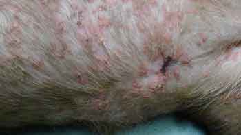
This is Daisy. She is thought to be a five month old female intact pit bull mix. She was found on the streets in South Central Los Angeles by a coworker of the current owner who works for the L.A. Police Department (not animal control). The coworker could not keep Daisy, so that is how Sue came to adopt Daisy. Sue and Marie have three children and two other rescued dogs.
Initially, Daisy had just a few lesions on her back, but later the lesions started spreading and getting worse. She was treated with antibiotics but not making improvement so she was referred to Animal Dermatology Clinic. Skin biopsies showed evidence of third-degree burns with secondary infection; possibly a caustic substance (acid or hot liquid) was poured on her back. Daisy’s skin has responded to some conservative management with oral medications. However, the degree of contraction caused by healing tissue and risks of serious infection make more aggressive therapy necessary to give her the best prognosis.
Animal Dermatology Clinic is working closely with Veterinary Surgical Specialists (Tustin, CA) to get Daisy the proper medical attention and care. Animal Assistance League of Orange County is accepting donations for Daisy’s medical care at their website
www.aaloc.com